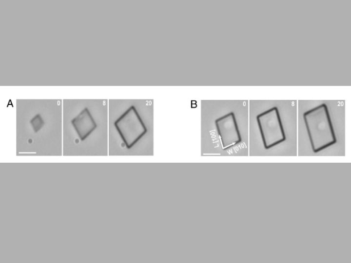
University of Houston video — the first of its kind — shows how cholesterol crystals grow inside a tiny fluid device using a mix of alcohol and water. Over 20 minutes, a thin crystal forms as cholesterol builds up.


A pair of University of Houston professors, known globally for their seminal contributions to crystal engineering with specific breakthroughs in the design of therapeutics to prevent crystallization in human diseases, are discovering how cholesterol crystals are formed in environments that mimic the human body.
Jeffrey Rimer, Abraham E. Dukler Endowed Professor of Chemical Engineering, and Peter Vekilov, Frank L. Worley Endowed Professor of Chemical Engineering, have published these findings and the corresponding videos of surface growth, which offer valuable insights into cholesterol's role in heart disease, in Proceedings of the National Academies of Science.
It’s the first time anyone has been able to take images of the surface growth of cholesterol crystals in real time at near molecular resolution.
Cholesterol crystals play an important role in diseases like heart disease and gallstones. The crystals can build up in blood vessels or the gallbladder, causing blockages or pain and ultimately disease, yet relatively few studies have explored the specific processes behind how cholesterol forms crystals, even though the stakes are high.
“These insights provide a foundation for future design of modifiers that selectively interact with crystal surfaces to cooperatively enhance growth inhibition, thus generating new opportunities to discover therapeutics that improve human health by counteracting the deleterious effects associated with cholesterol precipitation,” said Rimer.
The findings
Rimer, Vekilov and team found that crystals grow in layers, and the layers interact with each other to slow down the growth of the crystal. This causes the crystal to grow more slowly in certain areas compared to others.
But first, the team identified a special solvent that mimics the body's natural environment. This provided the setting for creating cholesterol crystals with the correct, physiologically relevant structure, allowing the scientists to watch how they grow in real time.
“Using a binary mixture of water and isopropanol, with the latter serving as a surrogate for lipids in physiological environments, we show that cholesterol monohydrate crystals grow classically by the nucleation and spreading of new crystal layers,” said Vekilov.
The team took pictures over time to watch how the cholesterol crystals grow. They saw them growing in layers and watched as these layers spread across the surface of the crystal and then join other growing layers on the edges.
“Time-resolved imaging confirms that layers are generated by dislocations and monomers incorporate into advancing steps after diffusion along the crystal surface and not directly from the solution,” said Rimer.
“This finding stands in contrast to numerous other systems, in which classical mechanisms lead to unhindered growth by spreading of single layers.”